Dr. Makda provided EXCEPTIONAL care to my father for his knee replacements.
Dr. Makda provided EXCEPTIONAL care to my father for his knee replacements. His attention to his patients and willingness to explain the treatment pla...
The knee is a complex joint made up of different structures including bones, tendons, ligaments, cartilage, and muscles. They all work together to maintain normal function and provide stability to the knee during movement.
The knee can be described as a hinge joint, similar to a door hinge. The knee not only bends back and forth but also rotates with movement.
Having a well-functioning healthy knee is essential for our mobility and ability to participate in various activities. Understanding the anatomy of the knee enhances your ability to discuss and choose the right treatment procedure for knee problems with your doctor.
The knee is a hinge joint made up of three bones, the thighbone (femur), the shinbone (tibia), and the knee cap (patella).
The knee is divided into 3 compartments:
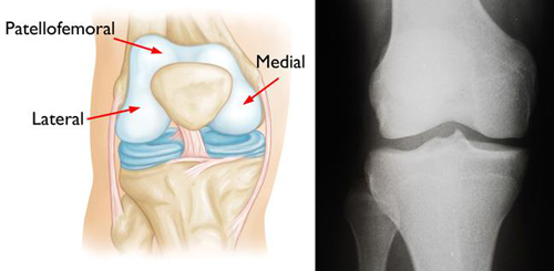
(Left) A normal knee joint: The medial, lateral, and patellofemoral compartments are shown with red arrows. (Right) An x-ray of a normal knee joint showing healthy space between the bones.
Image Credit: OrthoInfo
Movement of the knee joint causes friction between the three bones. To reduce this friction, the bones involved in movement are covered with a white, shiny, slippery layer called cartilage. The cartilage provides a smooth surface that facilitates easy movement. Cartilage rubbing against articular cartilage in a joint is ten times smoother than ice rubbing against ice!
Within the knee joint, between the femur and tibia, there are two C shaped structures called menisci. Menisci function to provide stability to the knee by spreading the weight of the upper body across the entire surface and preventing the weight from concentrating onto a small area, which could damage the articular cartilage. The menisci also act as a cushion or “donut” between the femur and tibia by absorbing the shock produced by activities such as walking, running and jumping.
To further reduce friction between the surfaces of the bones, the knee joint produces a thick clear fluid called synovial fluid. This joint fluid lubricates and nourishes the cartilage and allows the bones to move freely and smoothly.
Ligaments are tough bands of tissue that connect one bone to another bone. The ligaments of the knee function to stabilize the knee joint. There are two important groups of ligaments that hold the bones of the knee joint together, collateral ligaments and the cruciate ligament.
Collateral ligaments are present on either side of the knee. They function to prevent the knee from moving too far during side to side motion. The collateral ligament on the inside is called the medial collateral ligament (MCL) and the collateral ligament on the outside is called the lateral collateral ligament (LCL).
Cruciate ligaments, present inside the knee joint, control the back-and-forth motion of the knee. The cruciate ligament in the front of the knee is called anterior cruciate ligament or ACL and the cruciate ligament in the back of the knee is called posterior cruciate ligament or PCL.
There are two major muscles, the quadriceps and the hamstrings, which enable movement of the knee joint. The quadriceps muscles are in the front of the thigh. When the quadriceps muscles contract, the knee straightens. The hamstrings are in the back of the thigh. When the hamstring muscles contract, the knee bends.
Dr. Makda provided EXCEPTIONAL care to my father for his knee replacements.
Dr. Makda provided EXCEPTIONAL care to my father for his knee replacements. His attention to his patients and willingness to explain the treatment pla...
I had needed knee replacements for quite awhile.
I had needed knee replacements for quite awhile. I even saw other doctors about it but never moved forward as I realized now it all was never explaine...
Dr. Makda is wonderful. He is kind, very knowledgeable and offers different types of recovery for knee problems. After 3 visits I was released from ca...
My left knee was replaced in the hands of Dr Makda last July
My left knee was replaced in the hands of Dr Makda last July. Now my right hip in 2 weeks (mid April 2019).
All went as expected. Have to do all the therapy if you want to recover fast.
All went as expected. Have to do all the therapy if you want to recover fast. Very happy with my new left knee.
100 Village Green Drive
Suite 120
Lincolnshire, IL 60069